
GEORGIAN
BIOMEDICAL
& MEDICAL NEXUS
Former Georgian Biomedical News
ISSN (Online): 2720-8796
CLINICAL CASE
Successful Urgent Surgical Repair of an Intact 11.2-cm Ascending Aortic Aneurysm with Severe Secondary Aortic Regurgitation: A Case Report
Lasha Basiladze1, Simon Matikashvili1,ID, Levan Chelidze1,ID, Lasha Tabagari1, Irakli Shurgaia1,
Levan Totladze1, Ana Kereselidze1
ABSTRACT
Giant ascending aortic aneurysms (>10 cm) are uncommon because most thoracic aortic aneurysms are detected or become complicated before reaching this size. Nonspecific cardiopulmonary symptoms and mass effect may dominate their clinical presentation.
A 44-year-old man with a 2-month history of progressive retrosternal discomfort, dyspnea, fatigue, and reduced exercise tolerance was transferred urgently after an acute worsening of chest pain and dyspnea. He had previously been treated empirically for presumed atypical pneumonia without improvement. Computed tomography angiography demonstrated an intact, non-dissected proximal ascending aortic aneurysm measuring 10.7×11.2 cm, without contrast extravasation or periaortic hematoma. Transthoracic echocardiography demonstrated severe secondary aortic regurgitation, marked left ventricular dilation (LVEDD 7.0 cm; LVEDV 189 mL), and an LVEF of 45%.
After blood-pressure control and hemodynamic stabilization, the patient underwent urgent composite aortic root/ascending aortic replacement using a Bentall technique with a 25-mm mechanical bileaflet valve and a 30-mm woven Dacron graft. Myocardial arrest was achieved with ostial Del Nido cardioplegia.
The patient was extubated 4 hours postoperatively. At 3 months, he reported complete resolution of dyspnea and chest pressure and was in NYHA functional class I. Follow-up CTA demonstrated stable graft reconstruction without perigraft collection, pseudoaneurysm, or prosthetic dehiscence.
This case illustrates that an intact giant ascending aortic aneurysm may present with misleading respiratory symptoms and severe secondary aortic regurgitation. Prompt cross-
sectional imaging and definitive surgery can achieve a favorable early outcome even in extreme aortic dilation. No causal inference regarding the superiority of a specific cardioplegia strategy can be made from a single case.
Keywords: Aortic regurgitation; Ascending aortic aneurysm; Bentall procedure; Del Nido cardioplegia.; Giant aneurysm.
DOI: 10.52340/GBMN.2026.01.01.182
INTRODUCTION
Thoracic aortic aneurysm is commonly described as “giant” when its maximal diameter exceeds approximately 10 cm. Such dimensions are rarely encountered because the probability of rupture, dissection, and death rises substantially with increasing aortic size. In classic natural-history data, thoracic aortic aneurysms >6 cm were associated with annual rates of rupture of 3.7%, rupture or dissection of 6.9%, death of 11.8%, and a composite of death, rupture, or dissection of 15.6%. 1,2,5 Contemporary ACC/AHA and ESC guidance recommends prophylactic intervention at substantially smaller diameters in appropriate patients, with thresholds modified by anatomy, growth rate, family history, genetic disease, body size, and operative expertise. 3,4
The mechanical relationship described by Laplace’s law (wall stress proportional to pressure and radius and inversely proportional to wall thickness) predicts that, with pressure and wall thickness held constant, circumferential wall stress increases approximately linearly as radius increases - not exponentially. In vivo behavior is more complex because wall thickness, geometry, extracellular matrix, and tissue material properties also change during aneurysmal remodeling.
Intact giant ascending aortic aneurysms have nevertheless been reported, including lesions of 11-14 cm and larger, often with compressive symptoms and technically demanding surgical repair. 6-10 The present case is notable for the combination of young age, extreme intact aneurysmal dilation, severe secondary aortic regurgitation with left ventricular remodeling, a pulmonary-type clinical presentation with delayed diagnosis, and successful urgent surgical reconstruction.
CASE
Patient information and initial presentation
On May 19, 2026, a 44-year-old man was transferred by specialized emergency medical transport from a regional medical center to a tertiary cardiovascular institute for urgent evaluation. On arrival, he had acute dyspnea, exertion-related shortness of breath, non-radiating anterior chest pain, and a blood pressure of 160/90 mmHg. A single admission blood-pressure value was therefore documented as elevated; the available record did not establish chronic severe hypertension.
The patient described a 2-month history of progressive retrosternal discomfort, dyspnea, generalized fatigue, and reduced exercise tolerance. He had been treated as an outpatient for presumed atypical community-acquired pneumonia without clinical improvement. A sudden increase in chest pain and dyspnea on the morning of admission prompted emergency reassessment and transfer.
The documented past medical history was otherwise negative for diagnosed chronic disease. Social history included an approximately 40 pack-year smoking history and decades of heavy manual physical work. These exposures may contribute to vascular risk or transient hemodynamic loading, but they cannot by themselves establish the etiology of an aneurysm of this magnitude in a 44-year-old patient.
Physical examination
Neurological examination: The patient was conscious, alert, and fully oriented. Cranial nerves II-XII were intact; pupils were equal and reactive. Motor strength was 5/5 bilaterally, without focal neurological deficits or meningeal signs.
Cardiovascular examination: Heart sounds were markedly distant/muffled. Carotid, radial, femoral, and dorsalis pedis pulses were palpable, symmetric, and synchronous bilaterally, with no radio-femoral delay. No peripheral edema was documented.
Respiratory examination: Thoracic expansion was symmetric. Percussion demonstrated normal pulmonary resonance. Vesicular breath sounds were diminished bilaterally, most prominently in the right mid-to-lower lung fields.
Abdominal examination: The abdomen was soft, non-tender, and non-distended, without a palpable pulsatile mass or hepatosplenomegaly.
Diagnostic assessment
Computed tomography angiography (CTA): CTA demonstrated massive aneurysmal dilation of the proximal ascending thoracic aorta, with the abnormal segment beginning in the region of the sinotubular junction/proximal ascending aorta. The maximum cross-sectional diameter was 10.7×11.2 cm (FIG.1). The aortic arch and descending thoracic aorta were described as preserved in caliber (3.8 cm). No intimal flap, double lumen, intramural hematoma, contrast extravasation, or periaortic hematoma was identified, supporting an intact, non-dissected aneurysm. Segment-specific orthogonal measurements of the annulus, sinuses of Valsalva, sinotubular junction, and distal ascending aorta were not available in the source documentation.
FIGURE 1. Preoperative computed tomography angiography axial view of an aneurysmal dilation
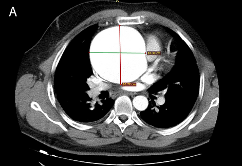
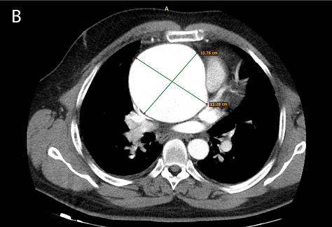
Explanations: A. Preoperative CTA axial image at the level of maximal aneurysmal dilation, demonstrating an ascending aortic diameter of approximately 10.7×11.2 cm without an intimal flap. B. Preoperative CTA axial image at a second level, demonstrating the giant ascending aortic aneurysm and associated displacement/mass effect on mediastinal structures.
Transthoracic echocardiography (TTE): TTE demonstrated marked proximal aortic dilation (reported maximal diameter 11.0 cm), severe secondary aortic regurgitation attributed to loss of leaflet coaptation in the setting of marked annular/root distortion, severe left ventricular dilation (LVEDD 7.0 cm; LVEDV 189 mL), and moderately reduced LVEF (45%). The left atrium measured 4.9 cm, the right ventricular dimension was 2.9 cm, and the inferior vena cava measured 1.8 cm with <50% inspiratory collapse. Quantitative aortic-regurgitation parameters (e.g., vena contracta, effective regurgitant orifice area, regurgitant volume/fraction, or holodiastolic flow reversal) were not available in the current record.Laboratory evaluation: Mild leukocytosis was present (WBC 12.93×10⁹/L). Anti-HCV, HIV, and Treponema pallidum testing were non-reactive. Renal function was preserved (serum creatinine 0.9 mg/dL; BUN 16 mg/dL), and the hepatic/metabolic panel was within reference limits.Etiologic considerationsGiven the patient’s age (44 years) and extreme proximal thoracic aortic dilation, contemporary guidance supports careful assessment for heritable thoracic aortic disease, bicuspid aortic valve- associated aortopathy, syndromic features, and relevant family history. 3,4 In the material available for this report, a three-generation family history, formal syndromic assessment, genetic testing, definitive native valve morphology (bicuspid versus tricuspid), and histopathologic characterization of the resected aortic wall were not documented. The absence of these data limits etiologic attribution. Tobacco exposure and possible episodic pressure loading from heavy manual work should therefore be considered potential modifiers rather than established causes. Clinical timeline
Transthoracic echocardiography (TTE): TTE demonstrated marked proximal aortic dilation (reported maximal diameter 11.0 cm), severe secondary aortic regurgitation attributed to loss of leaflet coaptation in the setting of marked annular/root distortion, severe left ventricular dilation (LVEDD 7.0 cm; LVEDV 189 mL), and moderately reduced LVEF (45%). The left atrium measured 4.9 cm, the right ventricular dimension was 2.9 cm, and the inferior vena cava measured 1.8 cm with <50% inspiratory collapse. Quantitative aortic-regurgitation parameters (e.g., vena contracta, effective regurgitant orifice area, regurgitant volume/fraction, or holodiastolic flow reversal) were not available in the current record.
Laboratory evaluation: Mild leukocytosis was present (WBC 12.93×10⁹/L). Anti-HCV, HIV, and Treponema pallidum testing were non-reactive. Renal function was preserved (serum creatinine 0.9 mg/dL; BUN 16 mg/dL), and the hepatic/metabolic panel was within reference limits.
Etiologic considerations
Given the patient’s age (44 years) and extreme proximal thoracic aortic dilation, contemporary guidance supports careful assessment for heritable thoracic aortic disease, bicuspid aortic valve- associated aortopathy, syndromic features, and relevant family history. 3,4 In the material available for this report, a three-generation family history, formal syndromic assessment, genetic testing, definitive native valve morphology (bicuspid versus tricuspid), and histopathologic characterization of the resected aortic wall were not documented. The absence of these data limits etiologic attribution. Tobacco exposure and possible episodic pressure loading from heavy manual work should therefore be considered potential modifiers rather than established causes.
Clinical timeline (TAB.2)

Abbreviations: BP, blood pressure; CTA, computed tomography angiography; LVEDD, left ventricular end-diastolic diameter; LVEF, left ventricular ejection fraction; NYHA, New York Heart Association; TTE, transthoracic echocardiography.
Surgical management
After blood-pressure control and hemodynamic stabilization, the patient underwent urgent operative repair during the index hospitalization.
Operative technique (Bentall procedure)
Access and cannulation: Median sternotomy was performed with particular caution because of the proximity of the aneurysmal wall to the posterior sternum. Cardiopulmonary bypass was established using right subclavian arterial cannulation and central dual-stage venous cannulation.
Myocardial protection: The aorta was cross-clamped proximal to the innominate artery. Cold hyperkalemic Del Nido cardioplegia was administered directly into the coronary ostia and produced electromechanical arrest. The available operative summary does not provide the total cardioplegia volume, exact formulation, cross-clamp duration, cardiopulmonary bypass time, or whether redosing was required.
Resection and reconstruction: The markedly dilated, thin-walled ascending aorta was resected. The native aortic valve was described intraoperatively as severely stretched and incompetent. A 25-mm mechanical bileaflet valve conduit incorporated into a 30-mm woven Dacron vascular graft was secured to the aortic annulus using interrupted pledgeted mattress sutures.
Coronary reimplantation and distal anastomosis: The left and right coronary ostia were mobilized as buttons and reimplanted into the Dacron conduit with continuous polypropylene sutures. The distal graft was anastomosed to the proximal aortic arch. The operative summary describes normothermic conditions and does not report the use of circulatory arrest.
Because the patient was young and the preoperative mechanism of regurgitation was described as secondary leaflet non-coaptation, the rationale for composite valve-graft replacement versus valve-sparing root replacement is clinically relevant. The documented severe leaflet stretching/incompetence and annular/root distortion support the choice of a Bentall-type reconstruction. However, detailed cusp morphology, annular measurements, and a formal valve-sparing assessment were not available in the current source record. These details should be added if available.
Postoperative course and follow-up
The procedure was completed without a reported technical or intraoperative adverse event. The patient was extubated 4 hours postoperatively on postoperative day 0 after satisfactory arterial blood-gas parameters and full recovery of consciousness. The source record does not provide postoperative echocardiographic measurements, cardiopulmonary bypass-related biomarkers, transfusion requirements, ICU length of stay, total hospital length of stay, or a detailed complication profile.
At the 3-month outpatient evaluation, the patient reported complete resolution of preoperative dyspnea and chest pressure and had NYHA class I functional capacity. Follow-up CTA demonstrated stable reconstruction. The 30-mm Dacron graft and mechanical valve prosthesis were described as being in satisfactory anatomical position, without perigraft fluid collection, pseudoaneurysm, or prosthetic dehiscence (FIG.2). Quantitative 3-month echocardiographic data (LVEF, LV dimensions/volumes, prosthetic gradients, and paravalvular regurgitation) and the anticoagulation regimen/INR target were not available in the current record.
FIGURE 2. Postoperative 3-month computed tomography angiography axial images


Explanations: A. Postoperative 3-month CTA axial image demonstrating the reconstructed proximal aorta/aortic root region after Bentall repair. B. Postoperative 3-month CTA axial image showing stable post-sternotomy and graft-related postoperative findings without evidence of sternal diastasis.
DISCUSSION
Clinical rarity and literature context
The principal value of this report is not aneurysm diameter alone. Giant ascending aortic aneurysms of 11–14 cm have been described previously (TAB.2). 6-10 Rather, the educational contribution lies in the combination of an intact 11.2-cm aneurysm in a relatively young patient, delayed recognition during treatment for presumed pulmonary infection, severe secondary aortic regurgitation with left ventricular remodeling, and successful urgent reconstruction.
The extreme size should be interpreted in the context of natural-history data and current guideline thresholds. Davies et al. reported that thoracic aneurysms >6 cm were associated with an annual rupture-or-dissection rate of approximately 6.9% and a composite annual rate of death, rupture, or dissection of 15.6%. 1 Current ACC/AHA and ESC recommendations support elective intervention at much smaller diameters in appropriate patients, particularly when additional high-risk features or heritable disease are present. 3,4 Thus, survival to an intact diameter of 11.2 cm represents unusual natural history, but not an unprecedented lesion.
TABLE 2. Representative published reports of giant ascending aortic aneurysms
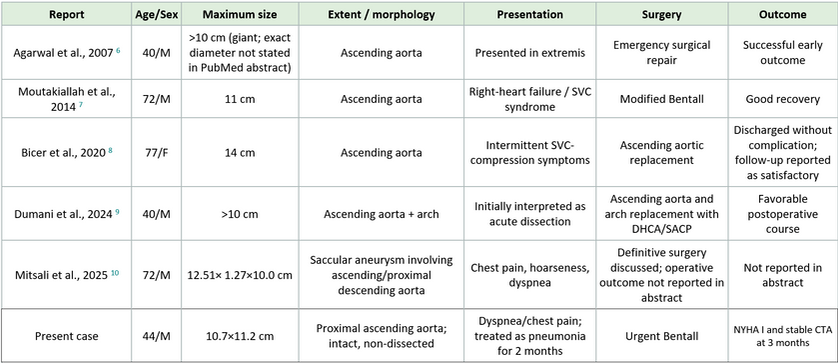
Abbreviations: CTA, computed tomography angiography; DHCA, deep hypothermic circulatory arrest; NYHA, New York Heart Association; SACP, selective antegrade cerebral perfusion; SVC, superior vena cava.
Etiologic assessment in a young patient
Extreme thoracic aortic disease at age 44 should prompt evaluation beyond common acquired cardiovascular risk factors. Contemporary guidelines emphasize assessment for heritable thoracic aortic disease, syndromic features, bicuspid aortic valve, and relevant family history, with genetic evaluation and family imaging when clinically indicated. 3,4 The present report cannot determine etiology because family-history details, formal syndromic/genetic assessment, native valve morphology, and histopathology were not available. Accordingly, previous language attributing the aneurysm primarily to smoking or manual labor has been removed. These factors may influence vascular risk and hemodynamic loading but do not establish causation.
Diagnostic pitfall and pulmonary-type presentation
Large thoracic aortic aneurysms can produce nonspecific respiratory symptoms through mediastinal mass effect and compression of adjacent structures. Published cases have described cough, dyspnea, hoarseness, tracheal compression, and superior vena cava syndrome. 7,8,10 In the present patient, persistent dyspnea and chest discomfort had been treated as atypical pneumonia for approximately 2 months. The key diagnostic lesson is therefore not that all persistent respiratory symptoms mandate CTA, but that unexplained or refractory cardiopulmonary symptoms-especially when accompanied by abnormal cardiovascular examination, mediastinal widening, aortic-regurgitation findings, pulse abnormalities, or concern for acute aortic disease-should prompt consideration of thoracic aortic pathology and appropriate cross-sectional imaging.
Surgical strategy and valve choice
The Bentall procedure remains an established strategy when the aortic root/annulus and valve require replacement. Valve-sparing root replacement may be appropriate in selected patients when the native valve is suitable, and the procedure can be performed by an experienced aortic team. 3 In this case, the operative record described markedly stretched and incompetent leaflets in the setting of severe annular/root distortion; however, detailed cusp morphology and a formal valve-sparing feasibility assessment were not documented. Providing these data would strengthen the rationale for mechanical composite graft replacement in this 44-year-old patient.
Myocardial protection
Del Nido cardioplegia has been used increasingly in adult cardiac surgery and has been evaluated in randomized and observational studies. 11,12 In this patient, a single ostial dose achieved satisfactory myocardial arrest and the early postoperative course was uncomplicated. However, this single case cannot establish that Del Nido cardioplegia caused the early extubation or was superior to alternative cardioplegia strategies. Interpretation would also be strengthened by reporting the exact local formulation, delivered volume, cardiopulmonary bypass time, cross-clamp time, and whether redosing was required.
Limitations
This report has several limitations. First, the available documentation does not include a complete family history, formal syndromic/genetic evaluation, native aortic valve morphology, or histopathology of the resected aorta; therefore, the etiology cannot be established. Second, segment-specific CTA measurements of the aortic root and quantitative echocardiographic grading of aortic regurgitation were unavailable. Third, key operative variables - including cardiopulmonary bypass time, aortic cross-clamp time, detailed temperature/perfusion strategy, cardioplegia volume/formulation, transfusion burden, and device manufacturer/model - were not provided in the source material. Fourth, postoperative echocardiographic remodeling, prosthetic hemodynamics, anticoagulation details, and complete in-hospital morbidity data were unavailable. Finally, follow-up was limited to 3 months; therefore, only an early favorable outcome can be claimed.
CONCLUSIONS
An intact 11.2-cm ascending aortic aneurysm can present with misleading respiratory symptoms and severe secondary aortic regurgitation despite the absence of acute dissection or rupture. In a young patient with unexplained or refractory cardiopulmonary symptoms, clinicians should maintain awareness of thoracic aortic disease and use appropriate cross-sectional imaging when the clinical context raises suspicion. Urgent Bentall repair in this case produced a favorable early clinical and imaging outcome at 3 months. Longer follow-up and more complete etiologic, pathological, operative, and echocardiographic characterization are required before broader mechanistic or long-term conclusions can be drawn.
AUTHOR AFFILIATION
1 Tbilisi Heart Center, Tbilisi, Georgia
INFORMED CONSENT
Written informed consent was obtained from the patient for publication of this case report, including the relevant clinical information and diagnostic imaging. All potentially identifying information was anonymized to protect patient confidentiality.
ETHICAL COMPLIANCE
According to institutional policy, formal Ethics Committee approval was not required for this single-patient, anonymized case report. The report was prepared in accordance with institutional ethical and confidentiality standards and the principles of the Declaration of Helsinki.
REFERENCES
-
Davies RR, Goldstein LJ, Coady MA, Tittle SL, Rizzo JA, Kopf GS, Elefteriades JA. Yearly rupture or dissection rates for thoracic aortic aneurysms: simple prediction based on size. Ann Thorac Surg. 2002;73(1):17-27; discussion 27-28. doi:10.1016/S0003-4975(01)03236-2.
-
Elefteriades JA. Natural history of thoracic aortic aneurysms: indications for surgery, and surgical versus nonsurgical risks. Ann Thorac Surg. 2002;74(5):S1877-S1880. doi:10.1016/S0003-4975(02)04147-4.
-
Isselbacher EM, Preventza O, Black JH 3rd, et al. 2022 ACC/AHA Guideline for the Diagnosis and Management of Aortic Disease: A Report of the American Heart Association/American College of Cardiology Joint Committee on Clinical Practice Guidelines. Circulation. 2022;146(24):e334-e482. doi:10.1161/CIR.0000000000001106.
-
Mazzolai L, Teixido-Tura G, Lanzi S, et al. 2024 ESC Guidelines for the management of peripheral arterial and aortic diseases. Eur Heart J. 2024;45(36):3538-3700. doi:10.1093/eurheartj/ehae179.
-
Coady MA, Rizzo JA, Hammond GL, et al. What is the appropriate size criterion for resection of thoracic aortic aneurysms? J Thorac Cardiovasc Surg. 1997;113(3):476-491. doi:10.1016/S0022-5223(97)70360-X.
-
Agarwal V, Yaliwal C, Ofo E, Kolvekar S. Giant ascending aortic aneurysm--a case report and review. Heart Lung Circ. 2007;16(5):385-388. doi:10.1016/j.hlc.2006.09.009.
-
Moutakiallah Y, Ait Houssa M, Seghrouchni A, et al. A giant aneurysm of ascending aorta revealed by superior vena cava syndrome. Heart Lung Circ. 2014;23(12):e264-e266. doi:10.1016/j.hlc.2014.08.001.
-
Bicer M, Yuksel A, Kan II. The largest reported giant ascending aortic aneurysm presented with superior vena cava syndrome. Braz J Cardiovasc Surg. 2020;35(5):834-837. doi:10.21470/1678-9741-2019-0151.
-
Dumani S, Likaj E, Dibra L, Ibrahimi A, Baboci A. Giant aneurysm of ascending aorta and aortic arch: a report of a rare case. Cureus. 2024;16(4):e59393. doi:10.7759/cureus.59393.
-
Mitsali T, Dewi DK, Hilman. Giant ascending aortic aneurysm: a rare case report. Radiol Case Rep. 2025;20(6):2797-2801. doi:10.1016/j.radcr.2025.02.030.
-
Matte GS, del Nido PJ. History and use of del Nido cardioplegia solution at Boston Children’s Hospital. J Extra Corpor Technol. 2012;44(3):98-103.
-
Ad N, Holmes SD, Massimiano PS, Rongione AJ, Fornaresio LM, Fitzgerald D. The use of del Nido cardioplegia in adult cardiac surgery: a prospective randomized trial. J Thorac Cardiovasc Surg. 2018;155(3):1011-1018. doi:10.1016/j.jtcvs.2017.09.146.
-
Gagnier JJ, Kienle G, Altman DG, Moher D, Sox H, Riley D; CARE Group. The CARE guidelines: consensus-based clinical case reporting guideline development. J Med Case Rep. 2013;7:223. doi:10.1186/1752-1947-7-223.