
GEORGIAN
BIOMEDICAL
& MEDICAL NEXUS
Former Georgian Biomedical News
ISSN (Online): 2720-8796
ORIGINAL RESEARCH
Early Sacubitril/Valsartan Treatment Attenuates Time-of-Day Blood Pressure Variation and Neurohumoral Alterations in DOCA-Salt Hypertensive Rats
Davit Goloshvili1,ID, Maia Okujava1,ID, Nikoloz Gongadze1, Manana Ghonghadze1,ID, Ketevan Pachkoria1,ID, Khatia Bediashvili1
ABSTRACT
Background. Diurnal blood pressure (BP) variation is strongly associated with cardiovascular morbidity, endothelial dysfunction, and target-organ damage in hypertension. The renin–angiotensin–aldosterone system (RAAS), endothelin-1 (ET-1), and melatonin are important regulators of both vascular tone and circadian hemodynamic rhythms. This study investigated the effects of early sacubitril/valsartan treatment on time-of-day BP variation and neurohumoral alterations in DOCA-salt hypertensive rats.
Objectives. To evaluate the preventive and therapeutic impacts of sacubitril/valsartan on blood pressure fluctuations and related serum biomarkers at specific diurnal time points.
Methods. Forty male rats were randomly divided into four groups (n=10/group): control, DOCA-salt hypertensive, therapeutic sacubitril/valsartan treatment after hypertension induction, and early sacubitril/valsartan treatment initiated simultaneously with hypertension induction. Hypertension was induced by intraperitoneal administration of deoxycorticosterone acetate (DOCA; 25 mg/kg) combined with 1% NaCl and 0.2% KCl in drinking water for 4 weeks. Sacubitril/valsartan was administered orally at 30 mg/kg/day. Systolic BP (SBP), diastolic BP (DBP), and heart rate (HR) were assessed weekly and at three specific time points (09:00, 15:00, and 21:00). Serum levels of ET-1, angiotensin II (Ang II), and melatonin were determined using ELISA.
Results. DOCA-salt hypertension significantly increased SBP, DBP, and HR compared with controls (p-values determined via post hoc analysis; p<0.001), accompanied by marked elevations in ET-1 and Ang II levels and reduced melatonin concentrations. Untreated hypertensive rats exhibited significant alterations in time-of-day BP values, with a loss of the normal fluctuations seen in controls. Therapeutic administration of sacubitril/valsartan restored near-normal BP values (SBP: 123.2±10 mmHg; DBP: 59.2±5 mmHg), normalized circadian hemodynamic fluctuations, reduced ET-1 and Ang II concentrations, and restored melatonin levels. Early initiation of treatment exerted an even more pronounced protective effect, preventing the development of hypertension, minimizing diurnal BP variability, and attenuating cardiovascular dysfunction.
Conclusions. Early initiation of sacubitril/valsartan effectively prevents the development of DOCA-salt hypertension, modifies time-of-day hemodynamic variations at the measured points, and regulates serum endothelin-1, melatonin, and angiotensin II profiles. These findings suggest that early neurohumoral modulation with sacubitril/valsartan may confer significant cardioprotective benefits beyond conventional BP reduction, particularly by preserving circadian cardiovascular regulation.
Keywords: Angiotensin II; Arterial hypertension; Circadian rhythm; DOCA-salt hypertension; Endothelin-1; Melatonin; Sacubitril/valsartan.
DOI: 10.52340/GBMN.2026.01.01.178
BACKGROUND
Arterial hypertension is a major modifiable risk factor for cardiovascular disease. Hypertension is a cardiovascular disease characterized by elevated blood pressure, with systolic> 140 mmHg and diastolic> 90 mmHg. Hypertension can be divided into three stages (prehypertension, stage 1 hypertension, and stage 2 hypertension) and three levels of risk (low, moderate, and high). Hypertension-induced organ damage refers to structural or functional changes in the arterial system and/or organs caused by elevated blood pressure. 1 Hypertension affects 25–43% of the world's population aged 18 and older. 2 It is a major modifiable risk factor for cardiovascular mortality. Hypertension can be secondary, resulting from a variety of causes, but the majority of patients have primary hypertension without a secondary cause. Although many strategies for treating arterial hypertension are now available, 8–12% of patients with hypertension still have uncontrolled blood pressure despite treatment. 3 The short- and long-term regulation of blood pressure involves a comprehensive coordinated action of the cardiovascular, renal, nervous, and endocrine systems. Arterial hypertension, in turn, leads to stroke, myocardial infarction, heart failure, kidney damage, and many other health problems. As previously mentioned, despite many treatment options, one in five patients has uncontrolled arterial hypertension; therefore, hypertension management is a major challenge for modern public health. The prevalence and clinical impact of hypertension are increasing worldwide, and its control is challenging, particularly in low-income countries. 4
There are many classifications of arterial hypertension, but scientists agree that resistant hypertension is a particularly severe form of this disease. It was first described about 50 years ago and has been the subject of active research since then. Resistant hypertension is elevated blood pressure that does not respond to intensive drug treatment.
Recent epidemiological studies have reported a prevalence of resistant hypertension in hypertensive patients of 10%, which is characterized by an increased risk of cardiovascular disease. 5 In addition, studies have identified subgroups of patients with even higher morbidity and mortality, which are likely to require more intensive drug management. 6
Recently, particular attention has been paid to the circadian rhythm of blood pressure (BP), as nocturnal and morning BP elevations are independent risk factors for cardiovascular disease. The renin-angiotensin-aldosterone system (RAAS) is involved in the circadian rhythm of blood pressure, and RAAS inhibitors play a crucial role in regulating it. The renin-angiotensin system (RAAS) is one of the important systems that regulate blood pressure. It involves a complex system of hormones, proteins, and enzymes. In addition to the classical endocrine (circulating) RAAS, additional research has revealed autocrine and paracrine effects of the RAAS. In addition, it has been shown that the RAAS can be produced locally and act in various organs, including endothelial cells, the adrenal and pituitary glands, the testes, ovaries, kidneys, heart, and eyes. 7 Because systemic RAAS components cannot readily penetrate most brain regions due to the blood-brain barrier (BBB), the brain RAAS (b-RAAS) is of particular importance as a local RAAS. 8 Angiotensinogen, a precursor of the RAAS, is found in pineal glial cells, and pinealocytes contain AT1b receptors. 9 Angiotensin-converting enzyme (ACE) and chymase, but not renin, are part of the enzymatic cascade that produces Ang II, suggesting the existence of non-renin pathways. 10 Ang II produced locally in the pineal gland may affect the synthesis of melatonin, the main hormone of the pineal gland and considered a key modulator of circadian rhythms. 11 Angiotensin II, which is produced by glial cells from angiotensinogen, stimulates tryptophan hydroxylase, the rate-limiting enzyme in melatonin synthesis, by binding to pineal AT1b receptors. Angiotensin and melatonin may interact to regulate central circadian rhythms in the suprachiasmatic nucleus (SCN) or peripherally, which is important for cardiovascular organs. 12 Circulating angiotensin II activates central pathways that increase sympathetic activity, decrease baroreflex sensitivity, and increase vasopressin secretion. 13 However, in hypertension, circulating angiotensin II can cross the blood-brain barrier and integrate into the hypothalamus and brainstem. 14 Healthy individuals experience a 10–20% drop in blood pressure at night, which is considered normal. Individuals whose blood pressure does not change by at least 10% during sleep are considered "non-dippers." 15 This type of hypertension is associated with activation of the renin-angiotensin-aldosterone system (RAAS), 16 an increased risk of chronic kidney disease (CKD), 17 and adverse cardiovascular events. 18,19 The combination of high salt intake and hypertension is associated with a significantly increased risk of cardiovascular mortality. 18 Night shift work, which disrupts circadian rhythms, increases the risk of various diseases, including cancer and cardiovascular disease. 20 Circadian rhythm disruption in night shift workers has been shown to result in significant increases in systolic blood pressure (SBP), diastolic blood pressure (DBP), and C-reactive protein (CRP), indicating systemic inflammation. The effect of circadian rhythm disruption on cardiovascular risk factors in healthy individuals has also been investigated. 21 In individuals with disrupted circadian rhythms, elevated levels of CRP, interleukin-1, resistin, and tumor necrosis factor (TNF) have been shown. Elevated levels of interleukin-1, resistin, and TNF have been associated with the progression of hypertension through activation of inflammatory pathways and endothelial dysfunction. 22 These findings suggest a direct link between disruption of normal circadian rhythms and increased risk of cardiovascular disease, and studies have implicated angiotensin II and melatonin in modulating circadian rhythms. Melatonin, known as the sleep hormone and secreted by the pineal gland, significantly reduces nocturnal blood pressure and also promotes normal sleep in patients with essential hypertension. Plasma renin activity (PRA), angiotensin-converting enzyme (ACE) activity, angiotensin II (AngII), aldosterone, and thyroid hormone concentrations also influence the maintenance of the 24-hour rhythm of blood pressure. 23 Blood pressure decreases physiologically during the night, but each 5% increase above the normal range can increase mortality risk by approximately 20%. 24 Myocardial infarction (MI) is a life-threatening condition that can be complicated by arrhythmia, shock, or heart failure. There is a strong association between elevated blood pressure and myocardial infarction. Disruption of the circadian rhythm of blood pressure or chronically elevated blood pressure can lead to pathological changes in normal myocardial perfusion and increase the risk of myocardial infarction. 25
METHODS
The experiments were carried out on 40 male rats weighing 200.0-250.0 g. The animals were housed in a vivarium with a temperature regime of 23±1oC, 50±5% humidity and 12 h light - 12 h dark (lights on at 07:00, lights off at 19:00). In accordance with the principles of the International Guidelines for Free Access to Food and Water and the criteria established by the Ethical Council of the Tbilisi State Medical University (Approval No: 51/25.12.2020]. Animals were handled in compliance with international guidelines, allowed a 7-day acclimatization period, and randomized into groups using a computer-based random number generator. Outcome assessors were blinded to the treatment groups during data collection. The animals were randomly divided into four groups (n=10 per group): Group I received normal drinking water for 4 weeks. Group II (Hypertensive Group): Received intraperitoneal injections of 25 mg/kg DOCA twice weekly and drinking water supplemented with 1% NaCl and 0.2% KCl for 4 weeks. Group III (Therapeutic Treatment Group): Received the same 4-week DOCA-salt protocol as Group II, followed by 2 weeks of oral sacubitril/valsartan therapy (30 mg/day). Group IV (Early/Preventive Treatment Group): Received oral sacubitril/valsartan therapy (30 mg/day) initiated simultaneously with the DOCA-salt protocol and maintained throughout the entire 4-week study period. To monitor the dynamics of DOCA-salt hypertension development, systolic, diastolic, and mean arterial pressure were measured from the tails of non-anesthetized rats in all four groups in a special chamber every week with the tail-cuff method on a Neurobotic device, with computer support. For this purpose, the animals were placed in the chamber to adapt for 30 minutes, after which the pressure values were measured 5 times at 5-10 minute intervals and averaged. In addition, heart rate was measured with the aforementioned device. To determine the time-of-day variation in systemic BP and HR, parameters were evaluated at three time points: 09:00, 15:00, and 21:00. Drug dosing was administered daily at 08:00, exactly 1 hour prior to the 09:00 measurement. Following the 4-week protocol (or 6 weeks for Group III), at the corresponding hours of blood pressure acrophases (the time when the indicator reaches its maximum), rats were anesthetized with pentobarbital (65 mg/kg i.p.). Blood collection was strictly standardized across all groups and performed at exactly 15:00 under identical environmental light conditions to avoid time-of-day and light-induced confounding artifacts on melatonin levels. Blood samples were collected via a catheter in the carotid artery. Serum concentrations of Endothelin-1, Angiotensin II (Ang II), and Melatonin (MT) were determined using quantitative ELISA kits (Cusabio, USA) in accordance with manufacturer guidelines.
Statistical processing of data
Quantitative data are presented as mean ± standard error of the mean (SEM). Main and interaction effects for hemodynamic variables measured across multiple time points were analyzed using a two-way repeated-measures ANOVA (Factors: Group and Time-of-Day). Serum biomarker levels were analyzed using a standard two-way ANOVA. Post hoc pairwise comparisons were performed using Tukey's adjustment for multiple testing. Normality and homogeneity of variances were verified via Shapiro-Wilk and Levene's tests, respectively. Statistical significance was defined as p<0.05, with exact p-values reported where applicable.
RESULTS
In the control group, the baseline SBP was 123.0±5.23 mmHg at 15:00. Following the 4-week administration of DOCA and high-salt water, SBP significantly increased, reaching 183.6±15.42 mmHg at 15:00. In Group III, therapeutic oral administration of sacubitril/valsartan (30 mg/day) reversed this hypertensive state, returning SBP values to near-control levels (123.2±10.84 mmHg). In Group IV, early co-administration of sacubitril/valsartan completely prevented the development of DOCA-salt hypertension, maintaining SBP at a normal baseline of 103.7±4.57 mmHg. Diastolic blood pressure followed a similar pattern: Group I control levels (60.4±3.10 mmHg at 15:00) rose dramatically to 97.9±8.19 mmHg in Group II, then dropped to 59.2±4.85 mmHg following therapeutic intervention (Group III), and remained at 51.8±609 mmHg under early preventive care (Group IV). (FIG.1); Mean HR was elevated in Group II (438.5±25.63 beats/min) relative to Group I (361.4±24.91 beats/min) but normalized effectively under therapeutic (370.2±31.00 beats/min) and preventive (371.1±15.67 beats/min) regimens (FIG.2).
FIGURE 1. Blood pressure values in the control and experimental groups

Abbreviations: DBP, diastolic blood pressure; SBP, systolic blood pressure.
FIGURE 2. Heart rate variability in control and experimental groups

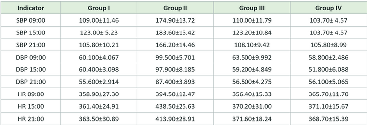
The average values of hemodynamic parameters in animals of all four study groups, namely systolic and diastolic blood pressures and heart rate, differed depending on the time of day. In particular, in animals of the control group, systolic blood pressure at 09:00 was 109±11 mmHg, at 15:00 it was 123.2±0 mmHg, and at 21:00 the minimum average value of systolic blood pressure was observed in this group, 105.8±10 mmHg (p<0.005). Also, in the same study group, different indicators were observed for diastolic blood pressure: at 09:00, it was 60.1±4 mmHg; at 15:00, 60.4±3 mmHg; and at 9:00 p.m., 55.6±2 mmHg (p<0.05). As for the heart rate of the control group animals, at 09:00 it was 358.9±27 beats/min, at 15:00 it was 361.4±24 beats/min, and at 21:00 it was 363.5±30 beats/min (p<0.001). Also, different hemodynamic parameters were observed after the development of DOCA-salt arterial hypertension: the mean systolic blood pressure at 09:00 was 183.6±15 mmHg, at 15:00 it was 174.9±13 mmHg, and at 21:00 it was 166.2±14 mmHg (p<0.005). Diastolic blood pressure values were 99.5±5 mmHg in the morning, 97.9±8 mmHg in the afternoon, and 87.4±4 mmHg in the evening (p<0.05). As for the heart rate in the hypertensive group, it was 394.5±12 beats/min at 09:00, 438.5±26 beats/min at 15:00, and 413.9±29 beats/min at 21:00 (p<0.001). In experimental group III, after treatment with the sacubitril/valsartan combination, the following circadian rhythm of hemodynamic parameters was observed: systolic blood pressure at 09:00 was 110±12 mmHg, at 15:00 was 123.2±11 mmHg, and at 21:00 was 108.1±10 mmHg (p<0.005). The diastolic blood pressure at 09:00 was 63.5±10 mmHg, at 15:00 - 59.2±5 mmHg, and at 21:00 - 56.5±4 mmHg. (p<0.05). The heart rate of the animals in the same experimental group at 09:00 was 356.4±15 beats/min, at 15:00 it was 370.2±31 beats/min, and at 21:00 it was 371.6±18 beats/min (p<0.05). In the animals of the fourth experimental group, the systolic blood pressure in the morning and afternoon hours was 103.7±4 mmHg, and at 21:00 - 105.8±9 mmHg. In animals of the same group, diastolic blood pressure at 09:00 was 58.8±2 mmHg, at 15:00 it was 51.8±6 mmHg, and at 21:00 it was 56.1±5 mmHg. As for the average heart rate: 365.7±11 beats/minute at 09:00, 371.1±15 beats/minute at 15:00, and 368.7±15 beats/minute at 21:00. (TAB.1).
TABLE 1. Circadian fluctuation of hemodynamic parameters in rats of the control and experimental groups, measured at 09:00, 15:00, and 21:00
The average level of the vasoconstrictor substance ET-1 in the animals of the control group was 18.28±3.47 pg/ml. Along with the increase in hemodynamic parameters, its level increased significantly in the animals of the second group, reaching 41.64±17.2 pg/ml (p<0.001). After treatment with the sacubitril/valsartan combination, a significant decrease in endothelin-1 levels was observed, with values of 15.12±9.51 pg/ml in the animals of the third experimental group (p<0.001). In the animals of the fourth group, there was no increase in endothelin-1 levels, and the average level was 16.32±2.71 pg/ml (p<0.001). (FIG.3).
FIGURE 3. Blood pressure values in the control and experimental groups

The mean angiotensin-II levels in group I animals were 12.5±4 pg/ml, which increased 5-fold in hypertensive rats to 63±26 pg/ml (p<0.001). In group III animals, after administration of sacubitril/valsartan, the mean Ang-II level decreased significantly to 11.1±5 pg/ml (p<0.001). Compared with animals in experimental groups I and III, the mean angiotensin-II level in group IV was moderately increased to 20.4±6.7 pg/ml. (FIG.4).
FIGURE 4. Concentrations of angiotensin II (ANG-II) in different experimental groups

The mean melatonin level in the control group was 9.8±3.6 pg/ml, whereas in the second group of animals, which developed arterial hypertension, it was significantly lower at 3.89±1.65 pg/ml (p<0.005). After treatment with the sacubitril/valsartan combination (oral dose of 30 mg/kg), the melatonin level in the third experimental group was 9.3±2 pg/ml (p<0.005). In contrast, the fourth group of animals had a mean value of 5.7±1.2 pg/ml (p<0.005) (FIG.5).
FIGURE 5. Concentrations of melatonin (MT) in different experimental groups

DISCUSSION
The results of the present study demonstrate that sacubitril/valsartan administration effectively reduces blood pressure and heart rate in DOCA-salt-treated rats. This therapeutic efficacy was observed both when administered after the establishment of hypertension (therapeutic group) and when initiated concurrently with DOCA-salt exposure (preventive group). Notably, early initiation of sacubitril/valsartan had a more pronounced effect in mitigating elevations in systemic systolic and diastolic blood pressure, suggesting potential utility for early intervention in this experimental model. A primary focus of this study was evaluating how early versus late sacubitril/valsartan intervention influences diurnal hemodynamic variations. It must be explicitly noted as a study limitation that systemic BP and HR were recorded at only three specific time points (09:00, 15:00, and 21:00). Consequently, these data reflect discrete time-of-day variations rather than a full, continuous 24-hour circadian profile. They cannot be used to mathematically define definitive circadian parameters such as nocturnal dipping, amplitude, acrophase, or phase shifts.
In our experimental model, the control group showed steady fluctuations across the measured hours. Conversely, the untreated hypertensive group exhibited an altered pattern, with significantly elevated blood pressure values persisting across the morning and afternoon points. While these findings indicate a clear disruption in normal time-of-day blood pressure variations under DOCA-salt exposure, interpreting these patterns requires caution. Because rats are nocturnal, their physiological rest/activity cycles are reversed compared to humans'. In our facility, the 12 h light/12 h dark cycle was strictly controlled, with lights on at 07:00 and lights off at 19:00. Thus, the 09:00 and 15:00 measurements correspond to the animals' early- to mid-quiescent (sleep) phase.
In contrast, the 21:00 measurement occurs during their active (dark) phase. The high blood pressure values observed during the resting phases in Group II align with a marked reduction in circulating melatonin levels. In the therapeutic group, treatment with the sacubitril/valsartan combination reduced these fluctuations, bringing values closer to those of the controls. Interestingly, in the early intervention group (Group IV), the day-to-evening variation across the three measured points was minimized, with blood pressure remaining baseline-like throughout. This stabilization suggests that early intervention may alleviate early-phase hemodynamic surges within this model. However, definitive conclusions regarding the preservation of true circadian rhythm or a reduced risk of future clinical cardiovascular events cannot be drawn without continuous 24-hour telemetry or structural tissue evaluations, which were outside the scope of this study.
Unlike studies performed in heart failure or high-renin conditions, sacubitril/valsartan treatment in our DOCA-salt model was associated with reduced serum angiotensin II concentrations. The DOCA-salt model is characterized by suppression of systemic renin-angiotensin system activity; therefore, sacubitril-mediated enhancement of natriuretic peptide signaling may have further inhibited renin release and angiotensin II generation. In addition, differences in analytical methodology, as Ang II was quantified by ELISA in the present study rather than equilibrium LC-MS/MS, may contribute to discrepancies between studies. 26,27
Our biochemical analyses revealed a substantial increase in serum endothelin-1 (ET-1) and angiotensin II (Ang II) in the untreated hypertensive group, paired with a significant decrease in serum melatonin. ET-1 is a potent endothelium-derived vasoconstrictor peptide. In the DOCA-salt rat model, elevated ET-1 levels increase vascular tone by binding to endothelin A (ETA) receptors on vascular smooth muscle cells. This effect is upregulated in the vascular wall under mineralocorticoid excess. 28 Previous in vitro research has shown that Ang II can stimulate the expression and secretion of ET-1, 29 and that these two peptides can act synergistically to exacerbate vasoconstriction in mineralocorticoid-induced hypertension. 30
However, the marked elevation of serum Ang II in our DOCA-salt group and its subsequent reduction by sacubitril/valsartan require careful mechanistic contextualization. The standard DOCA-salt model is traditionally characterized as a high-volume, salt-sensitive, low-renin model of hypertension, where systemic circulating components of the renin-angiotensin-aldosterone system (RAAS) are heavily suppressed by negative feedback from fluid retention. The observed increase in circulating Ang II in our protocol might stem from local tissue-level RAAS overactivation—particularly in the kidneys, vasculature, or central nervous system—that can spill over into the systemic circulation with prolonged exposure. Additionally, valsartan is an AT1 receptor blocker, a class of drug that typically causes a compensatory upward reflex in circulating Ang II due to the interruption of the negative feedback loop. The lower serum Ang II levels in our treated groups suggest that sacubitril/valsartan does not uniformly suppress the RAAS. Instead, simultaneous neprilysin inhibition by sacubitril increases natriuretic peptide levels, which may counterregulate systemic fluid balance, reduce sympathetic outflow, and ultimately modulate overall Ang II production in this specific high-salt state. To fully substantiate these pathways, future investigations should evaluate pre-analytical handling, local tissue RAAS expressions, and explicit ELISA cross-reactivities.
In our study, sacubitril/valsartan treatment significantly lowered serum ET-1 concentrations in the therapeutic group and limited its elevation in the preventive group. In the early-stage intervention group, serum ET-1 concentrations were kept low, suggesting that early limitation of these vasoconstrictors may help prevent the development of severe hypertension.
Regarding melatonin regulation, this pineal hormone plays a vital role in cardiovascular homeostasis via its antioxidant capacities. Melatonin production is highly time-dependent and sensitive to light and neurohumoral factors. In our protocol, blood collection for all experimental groups was strictly standardized and performed at exactly 15:00 under identical environmental light conditions. This eliminates confounding variations caused by sampling animals at shifting clock times or under varying light conditions. Elevated Ang II levels accompanied the reduced serum melatonin observed in the untreated hypertensive group. While renal Ang II does not cross an intact blood-brain barrier, hypertensive pathology can disrupt barrier integrity, potentially allowing peripheral peptides to interface with central structures and impact pineal synthesis. Following sacubitril/valsartan administration, serum Ang II decreased, while melatonin levels concurrently increased. In the early treatment group, where the onset of hypertension was attenuated, melatonin concentrations remained close to control values.
Several limitations must be acknowledged when interpreting these data. First, because tail-cuff plethysmography was limited to three time points per day, continuous diurnal profiling has not been verified. Second, while the term cardiovascular protection' is conceptually relevant, this study did not directly measure functional endothelial parameters, cardiac remodeling, oxidative stress markers, or renal structural injury. Therefore, the protective assertions made here are strictly limited to the observed modifications of serum biomarkers (ET-1, Ang II, melatonin) and the modulation of time-of-day blood pressure values.
CONCLUSIONS
In this DOCA-salt rat model, sacubitril/valsartan reduced blood pressure and heart rate and was associated with lower serum ET-1 and Ang II concentrations and higher melatonin levels compared with untreated hypertensive animals. Early administration attenuated the development of hypertension and reduced time-of-day variation in hemodynamic parameters. Further studies using continuous 24-hour BP telemetry and more detailed mechanistic assessment are needed to confirm whether sacubitril/valsartan truly preserves circadian blood pressure regulation.
AUTHOR AFFILIATION
1 Department of Medical Pharmacology, Tbilisi State Medical University, Tbilisi, Georgia
REFERENCES
-
Unger T, Borghi C, Charchar F, Khan NA, Poulter NR, Prabhakaran D, Ramirez A, Schlaich M, Stergiou GS, Tomaszewski M, Wainford RD, Williams B, Schutte AE. 2020 International Society of Hypertension global hypertension practice guidelines. J Hypertens. 2020 Jun;38(6):982-1004. doi: 10.1097/HJH.0000000000002453. PMID: 32371787.
-
Huffman MD, Lloyd-Jones DM. Global Burden of Raised Blood Pressure: Coming Into Focus. JAMA. 2017 Jan 10;317(2):142-143. doi: 10.1001/jama.2016.19685. PMID: 28097343.
-
Nguyen TN, Chow CK. Global and national high blood pressure burden and control. Lancet. 2021 Sep 11;398(10304):932-933. doi: 10.1016/S0140-6736(21)01688-3. Epub 2021 Aug 24. PMID: 34450082.
-
Verdecchia P, Cavallini C, Angeli F. Advances in the Treatment Strategies in Hypertension: Present and Future. J Cardiovasc Dev Dis. 2022 Mar 3;9(3):72. doi: 10.3390/jcdd9030072. PMID: 35323620; PMCID: PMC8949859.
-
Lamirault G, Artifoni M, Daniel M, Barber-Chamoux N, Nantes University Hospital Working Group On Hypertension. Resistant Hypertension: Novel Insights. Curr Hypertens Rev. 2020;16(1):61-72. doi: 10.2174/1573402115666191011111402. PMID: 31622203.
-
Chia R, Pandey A, Vongpatanasin W. Resistant hypertension-defining the scope of the problem. Prog Cardiovasc Dis. 2020 Jan-Feb;63(1):46-50. doi: 10.1016/j.pcad.2019.12.006. Epub 2019 Dec 19. PMID: 31863785; PMCID: PMC8272692.
-
Vajapey R, Rini D, Walston J, Abadir P. The impact of age-related dysregulation of the angiotensin system on mitochondrial redox balance. Front Physiol. 2014 Nov 24;5:439. doi: 10.3389/fphys.2014.00439. PMID: 25505418; PMCID: PMC4241834.
-
Triebel H, Castrop H. The renin angiotensin aldosterone system. Pflugers Arch. 2024 May;476(5):705-713. doi: 10.1007/s00424-024-02908-1. Epub 2024 Jan 17. PMID: 38233636; PMCID: PMC11033231.
-
Wright JW, Harding JW. The brain renin-angiotensin system: a diversity of functions and implications for CNS diseases. Pflugers Arch. 2013 Jan;465(1):133-51. doi: 10.1007/s00424-012-1102-2. Epub 2012 Apr 26. PMID: 22535332.
-
O. Baltatu, A. Lippoldt, A. Hansson, D. Ganten, and M. Bader, “Local renin-angiotensin system in the pineal gland,” Molecular Brain Research, vol. 54, no. 2, pp. 237–242, 1998.
-
Campos LA, Cipolla-Neto J, Amaral FG, Michelini LC, Bader M, Baltatu OC. The Angiotensin-melatonin axis. Int J Hypertens. 2013;2013:521783. doi: 10.1155/2013/521783. Epub 2013 Jan 8. PMID: 23365722; PMCID: PMC3556444.
-
Baltatu O, Afeche SC, José dos Santos SH, Campos LA, Barbosa R, Michelini LC, Bader M, Cipolla-Neto J. Locally synthesized angiotensin modulates pineal melatonin generation. J Neurochem. 2002 Jan;80(2):328-34. doi: 10.1046/j.0022-3042.2001.00701.x. PMID: 11902123.
-
Veerasingham SJ, Raizada MK. Brain renin-angiotensin system dysfunction in hypertension: recent advances and perspectives. Br J Pharmacol. 2003 May;139(2):191-202. doi: 10.1038/sj.bjp.0705262. PMID: 12770924; PMCID: PMC1573858.
-
Biancardi VC, Son SJ, Ahmadi S, Filosa JA, Stern JE. Circulating angiotensin II gains access to the hypothalamus and brain stem during hypertension via breakdown of the blood-brain barrier. Hypertension. 2014 Mar;63(3):572-9. doi: 10.1161/HYPERTENSIONAHA.113.01743. Epub 2013 Dec 16. PMID: 24343120; PMCID: PMC4080808.
-
Aksit E, Gursul E, Aydin F, Samsa M, Ozcelik F. Non-dipper hypertension is associated with slow coronary flow among hypertensives with normal coronary angiogram. Cardiovasc J Afr. 2017 Jan/Feb 23;28(1):14-18. doi: 10.5830/CVJA-2016-045. Epub 2016 May 13. PMID: 27177043; PMCID: PMC5423430.
-
Pati P, Fulton DJ, Bagi Z, Chen F, Wang Y, Kitchens J, Cassis LA, Stepp DW, Rudic RD. Low-Salt Diet and Circadian Dysfunction Synergize to Induce Angiotensin II-Dependent Hypertension in Mice. Hypertension. 2016 Mar;67(3):661-8. doi: 10.1161/HYPERTENSIONAHA.115.06194. Epub 2016 Jan 18. PMID: 26781276; PMCID: PMC4752410.
-
Hermida RC, Ayala DE, Mojón A, Fernández JR. Influence of circadian time of hypertension treatment on cardiovascular risk: results of the MAPEC study. Chronobiol Int. 2010 Sep;27(8):1629-51. doi: 10.3109/07420528.2010.510230. PMID: 20854139.
-
Kong F, Liu Q, Zhou Q, Xiao P, Bai Y, Wu T, Xia L. Dietary salt intake and cardiovascular outcomes: an umbrella review of meta-analyses and dose-response evidence. Ann Med. 2025Dec;57(1):2582065.doi:10.1080/07853890.2025.2582065. Epub 2025 Nov 16. PMID: 41243115; PMCID: PMC12624901.
-
de la Sierra A, Gorostidi M, Banegas JR, Segura J, de la Cruz JJ, Ruilope LM. Nocturnal hypertension or nondipping: which is better associated with the cardiovascular risk profile? Am J Hypertens. 2014 May;27(5):680-7. doi: 10.1093/ajh/hpt175. Epub 2013 Sep 23. PMID: 24061070.
-
Ohlander J, Keskin MC, Stork J, Radon K. Shift work and hypertension: Prevalence and analysis of disease pathways in a German car manufacturing company. Am J Ind Med. 2015 May;58(5):549-60. doi: 10.1002/ajim.22437. Epub 2015 Mar 13. PMID: 25773725.
-
Morris CJ, Purvis TE, Hu K, Scheer FA. Circadian misalignment increases cardiovascular disease risk factors in humans. Proc Natl Acad Sci U S A. 2016 Mar 8;113(10):E1402-11. doi: 10.1073/pnas.1516953113. Epub 2016 Feb 8. PMID: 26858430; PMCID: PMC4790999.
-
Fang C, Lei J, Zhou SX, Zhang YL, Yuan GY, Wang JF. Association of higher resistin levels with inflammatory activation and endothelial dysfunction in patients with essential hypertension. Chin Med J (Engl). 2013 Feb;126(4):646-9. PMID: 23422182.
-
Veglio F., Pietrandrea R., Ossola M., Vignani A., Angeli A. Circadian rhythm of the angiotensin converting enzyme (ACE) activity in serum of healthy adult subjects. Chronobiologia. 1987;14:21–25.
-
Ohkubo T., Hozawa A., Yamaguchi J., Kikuya M., Ohmori K., Michimata M., Matsubara M., Hashimoto J., Hoshi H., Araki T., et al. Prognostic significance of the nocturnal decline in blood pressure in individuals with and without high 24-h blood pressure: The Ohasama study. J. Hypertens. 2002;20:2183–2189. doi: 10.1097/00004872-200211000-00017.
-
Virag J.A., Lust R.M. Circadian influences on myocardial infarction. Front. Physiol. 2014;5:422. doi: 10.3389/fphys.2014.00422.
-
Rajzer P, Biegus J. Sacubitril/valsartan in a wide spectrum of heart failure patients (from mechanisms of action to outcomes in specific populations). Heart Fail Rev. 2025 Mar;30(2):387-405. doi: 10.1007/s10741-024-10471-1. Epub 2025 Jan 7. PMID: 39776087; PMCID: PMC11802626.
-
Polina I, Domondon M, Fox R, Sudarikova AV, Troncoso M, Vasileva VY, Kashyrina Y, Gooz MB, Schibalski RS, DeLeon-Pennell KY, Fitzgibbon WR, Ilatovskaya DV. Differential effects of low-dose sacubitril and/or valsartan on renal disease in salt-sensitive hypertension. Am J Physiol Renal Physiol. 2020 Jul 1;319(1):F63-F75. doi: 10.1152/ajprenal.00125.2020. Epub 2020 May 28. PMID: 32463726; PMCID: PMC7468826.
-
Schiffrin EL, Larivière R, Li JS, Sventek P. Enhanced expression of the endothelin-1 gene in blood vessels of DOCA-salt hypertensive rats: correlation with vascular structure. J Vasc Res. 1996 May-Jun;33(3):235-48. doi: 10.1159/000159151. PMID: 8924521.
-
Schiffrin EL. Role of endothelin-1 in hypertension and vascular disease. Am J Hypertens. 2001 Jun;14(6 Pt 2):83S-89S. doi: 10.1016/s0895-7061(01)02074-x. PMID: 11411770.
-
Lin YJ, Kwok CF, Juan CC, Hsu YP, Shih KC, Chen CC, Ho LT. Angiotensin II enhances endothelin-1-induced vasoconstriction through upregulating endothelin type A receptor. Biochem Biophys Res Commun. 2014 Aug 22;451(2):263 doi: 10.1016/j.bbrc.2014.07.119. Epub 2014 Aug 1. PMID: 25088996.